Fractures
Published: 27/01/2021
My summary notes on fracture classification, their clinical features and repair process (for compact and cancellous bone).
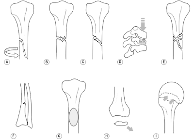
Classification
|
Fracture type |
Characteristics |
|
Open
or compound |
Bone
end or some other object has pierced the skin |
|
Closed |
Skin remains intact |
|
Displaced
or undisplaced |
Refers
to the alignment of the fractured bones |
|
Spiral or torsion |
A fracture where the bone has been twisted apart |
|
Transverse |
A
fracture in which the break is across a bone at a right angle to the long
axis of the bone |
|
Oblique |
A fracture in which the line of break runs obliquely (= slanting) to
the axis of the bone |
|
Compression
(or crush) |
Occurs
when the bone collapses e.g. vertebrae |
|
Comminuted |
Break or splinter of the bone into more than two fragments (common in
high-impact trauma) |
|
Greenstick |
A
fracture in which one side of a bone is broken and the other is bent |
|
Avulsion (or sprain) |
The detachment of a bone fragment that results from the pulling away
of a ligament, tendon, or joint capsule from its point of attachment on a
bone |
· Open (or compound) fracture can create a cause for concern due to the possibility of the introduction of microorganisms leading to osteomyelitis

Clinical features
·
Pain – marked tenderness around the site of the
fracture
·
Deformity
·
Oedema – localised immediately after injury and
becomes more extensive with time
·
Muscle spasm
·
Abnormal movement or crepitus – may be grating
between the broken ends of the bone
·
Loss of function
·
Hypovolaemic shock = an emergency condition in which
severe blood or fluid loss makes the heart unable to pump enough blood to the
body. This can cause many organs to stop working
Ø
fractured shaft may haemorrhage as much as three
pints
·
Limitation of joint movement – affected by
adhesion formation, tight muscles, pain, spasm, fear, mechanical obstruction or
swelling
·
Muscle atrophy
Repair process
Wolff’s law states that bone responds to the stresses
that are imposed upon it by rearranging its internal architecture to best
withstand the stresses
Simple summary – bone is laid down where it is needed and
reabsorbed where it is not
Repair process of compact bone
Haematoma
·
The haematoma forms due to the tearing of blood
vessels
·
Very small proportions of the bone immediately
adjacent to the fracture die and are gradually absorbed
Periosteal or endosteal proliferation
·
Proliferation of cells from the deep surface of
the periosteum adjacent to the fracture site
·
These cells are precursors to osteoblasts and
form around each fragment of bone
·
At the same time cells proliferate from the
endosteum in each fragment both types of cells forming tissue
·
This tissue gradually forms a bridge between the
bone ends
·
During this stage the haematoma is gradually
reabsorbed
Callus formation
·
Proliferating cells mature as osteoblasts or in
some instances as chondroblasts
·
Chondroblasts form cartilage
·
Osteoblasts lay down the intercellular matrix of
collagen and polysaccharide which becomes impregnated with calcium salts
forming immature bone called callus (or woven bone) [visible on X-ray]
Consolidation
·
Osteoblast activity results in the change of
primary callus to bone (which has a lamellar structure) at the end of this
stage union is complete
·
New bone forms a thickened mass at the fracture
site and obliterates the medullary cavity
Remodelling
·
The lamellar structure changes and the bone
adapts by strengthening along the lines of stress imposed upon it
·
The surplus bone formed during healing is
gradually removed and eventually the bone structure appears very similar to the
original
·
In children – healing is usually very good, difficult
to see fracture site on radiograph
·
In adults – may be a permanent area of
thickening which might be felt or seen in a superficial bone
Repair process of cancellous bone
·
Haematoma forms but these is no medullary cavity
·
Cancellous bone has a greater area of contact
between the fragments
·
Penetration of the bone-forming tissue is
facilitated by the open arrangement of trabeculae as it grows out from both
fragments
·
Osteogenic cells lay down intercellular matrix,
which calcifies to form woven bone
·
The process of remodelling then continues to
form the cancellous bone

Comments
Post a Comment